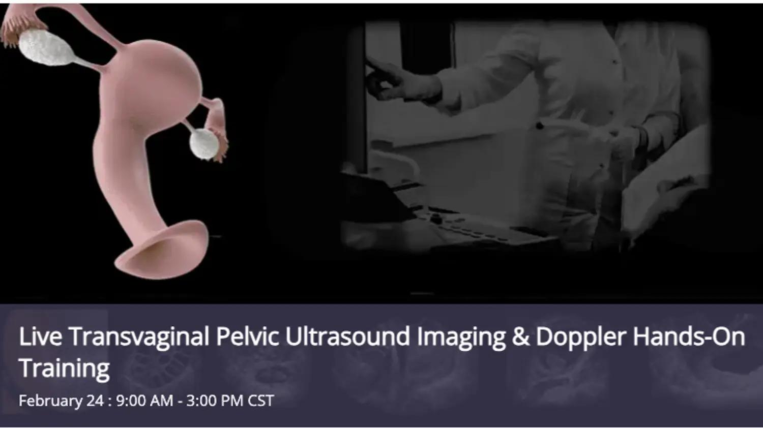
Live Transvaginal Pelvic Ultrasound Imaging & Doppler Hands-On Training
We’ve created the most effective live, hands-on transvaginal ultrasound training approach ever. You’ll learn the respectful approach to the patient and the rapid, authoritative protocol to survey, optimize, and document every pelvic structure.
We’ll build an absolutely precise protocol that ensures a complete ultrasound image and Doppler exam, that’s organized in sequential, scientific order. Likewise, we’ll explore all the machine’s options to optimize image and Doppler data, and connect the anatomic and physiologic ultrasound findings to normal and diseased states to help you enhance your patient’s care. Our personal post-class support stands by you in perpetuity for free.
You’ll Learn to Navigate the Pelvis with Competence and Confidence
This hands-on ultrasound training experience is much deeper than what your colleagues got in school; no matter where you’re starting with transvaginal imaging, we’ll take you farther.
A single day together will change your competence and confidence trajectory forever. You can spend months with your peers, observing, practicing, watching online, and still not grow as fast as you will with the live-learning skills we’ll impart.
Over forty years of experience in ultrasound practice, engineering, and teaching stand with you: both in Class and perpetuity.
Objectives:
Our approach is totally focused on the patient diagnosis. We are deeply familiar with virtually every ultrasound machine and the manufacturer’s rationale behind its design, features, and functions. No faculty members have any commercial interests or participation that might influence course content.
There is no formal test in this class: we evaluate you continuously and offer positive feedback and gentle corrections throughout. Upon completion of this activity, and through continued review, you should be able to:
- Demonstrate competence in inspecting the equipment for patient safety; cite the protocol for transducer sepsis before, during, and after the transcavitary exam
- Complete a systematic transvaginal ultrasound survey of the pelvis; identify, measure, and document all reproductive structures and related areas
- Identify, confirm, and locate the early implantation in suspected pregnancy
- Demonstrate proper technique to measure yolk sac and/or crown/rump length in the first trimester
- Note and communicate any focal rebound pain when reported by the patient
- Identify tubal pathology using image sonolocation and Doppler for physiologic assessment
- Extend the exam to completely survey the abdomen and posterior cul-de-sac when ectopic and/or heterotopic pregnancy is suspected
- Describe the protocol for translabial imaging
- Compare the spatial and input dynamic range sensitivity of the surface and intracavitary imaging probes; cite the finite limits of each
- Communicate a detailed summary of all findings using standardized terminology.
Topics:
The class is strictly small so we can spend time on the topics we need to cover and all the ones you want to discuss:
- How to intuitively orient yourself to the unconventional spatial representation posed by the transvaginal image display
- How to apply geometry to the precise pelvic protocol to confirm every structure and rule out every artifact
- How to sonolocate the ovary with authority every single time
- Color, power, and spectral Doppler’s findings matched to tissue physiology
- Confident evaluation of the posterior cut-de-sac; keys to differentiate serous fluid from hemorrhage
- The indications, role, and protocol form translabial imaging
- How and when to use surface pelvic imaging to evaluate the kidneys and urinary bladder
Who Will Benefit:
Allied Health Care Providers
This hands-on ultrasound training experience gives professionals from all specialties the solid foundation to broaden and deepen their talents and open doors to lifelong opportunities. These include Radiography and Respiratory Therapy Technologists, Traditional and Advanced-Practice Nurses, and PAs. Foreign Physicians who find it impractical to re-board in the USA or Canada have found this a valuable path to expressing their expertise.
This Course will not confer the requisite 12 months of clinical experience required to apply for credentialing, but it will give you a particular edge at the start, when your potential Director says, “Now let me see you scan….”
Acute Point of Care Providers: A Life-Saving Resource
Time is your currency. And the time you’ll save in deciding whether to irradiate your patient’s pelvis speaks to risk management in every way possible.
In the time it takes to ponder the benefit of ordering a TV pelvic exam and wait for the response & report, you’ll already have the critical finding decided.
There’s no secret to confidently locating the ovaries and inspecting the tubes, you just need to learn the protocol to define their sonolocation. And with mastery of color, power, and spectral Doppler you’ll have more technical knowledge than most Sonographers- and thus more authoritatively know how to validate their findings.
Sonography Students & Graduates:
Expand & Expound On Your Ultrasound Training
Students and Graduates of formal General Ultrasound programs will find far more job opportunities with echocardiography training on your resume. Many of our colleagues in this Class undertake it to build their foundation and/or expand upon it. Regardless of prior experience, we’ll be able to take you farther and deeper to prepare you for your future practice and credentialing. Your investment will pay out substantially when your potential Director says, “Let me see you scan.“